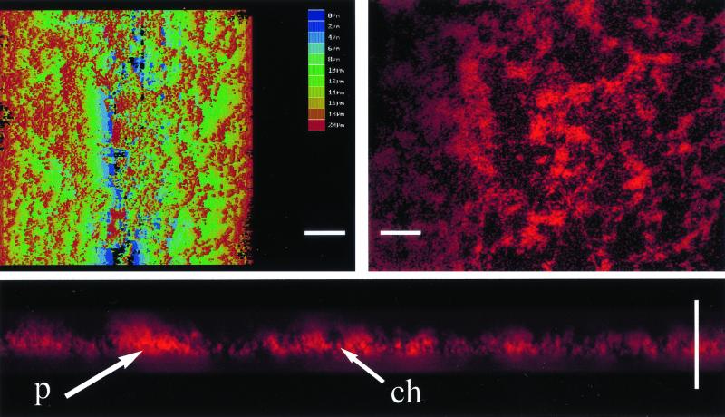
FIG. 2.
Laser confocal microscopy of biofilm produced by a csrA mutant. A topographical image of the biofilm formed by TR1-5MG1655 is shown in the left panel (white scale bar, 14 μm; virtual color code depicts biofilm height above the microscope slide, from 0 to 20 μm, in 2-μm increments), along with a 2-μm-thick cross-section at a depth of 6 μm in the right panel (scale bar, 10 μm) and a cross-section of a sagital view tilted (Q = 45°, F = 30°) in the bottom panel (scale bar, 20 μm), as visualized by confocal microscopy. Examples of apparent pillars (p) and channels (ch) are indicated.

