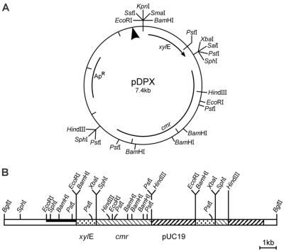
FIG. 1.
Physical maps of pDPX and pUCDV5. (A) Promoter probe pDPX. Restriction sites and the NarI-spanning illegitimate integration site (arrowhead) are given. (B) Plasmid pUCDV5. Parts of plasmid pDPX are duplicated in pUCDV5 because of a second, homologous recombination event that occurred after the first illegitimate integration; this second recombination was accompanied by an internal deletion of vector DNA. Relevant restriction sites are indicated. Open bar, R. fascians chromosomal DNA; black bar, probe. Locations of the promoterless xylE gene, cmr gene, and pUC19 vector DNA are shown.

