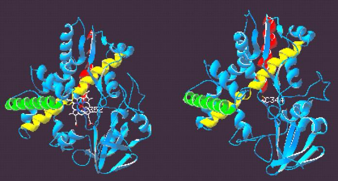
FIG. 3.
Ribbon diagrams of P450nor (left) and modeled three-dimensional structure of TxtC (right). Heme prosthetic group and cysteinyl thiolate ligand Cys352 are shown in P450nor, and the conserved putative ligand Cys344 is shown in TxtC. G, I, and L helices are depicted in green, yellow, and red, respectively. The figure was prepared with Swiss-PdbViewer v3.7b2.

