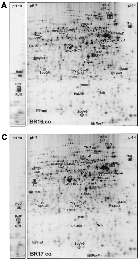
FIG. 3.
Differential protein synthesis patterns in B. subtilis wild type (BR16) versus a relA mutant (BR17) after norvaline treatment (indication of proteins belonging to the “RelA regulon”). 2D protein gels (autoradiograms) of l-[35S]methionine-labeled proteins (pH 4 to 7 and basic sections from pH 3 to 10) isolated from exponentially growing B. subtilis BR16 (A) and BR17 (C), from BR16 after 20 min of norvaline stress (0.05% [wt/vol]) (B), and from BR17 after 20 min of norvaline stress (0.05% [wt/vol]) (D) are shown. These autoradiograms show only proteins synthesized during the period of the l-[35S]methionine labeling. Proteins whose induction (circles) or repression (squares) is dependent on RelA are indicated. Proteins are indicated by arrows if the RelA dependence was only demonstrated by DNA macroarray analysis (see Table 1).


