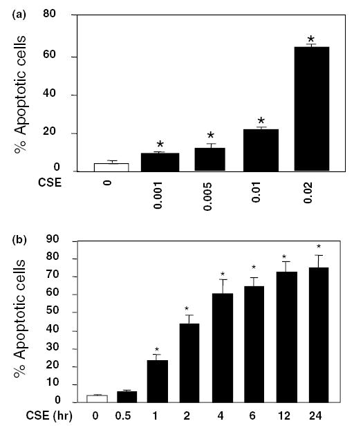
Fig. 1.

CSE induces apoptosis in HAECs. (a) Dose-dependent effects of CSE on endothelial apoptosis. Cells were treated with 0–0.02 U of CSE for 4 h and percentages of apoptotic cells (means + S.E.M., N = 3) were determined by FACS using TUNEL labeling. (b) Time course of HAEC apoptosis induced by 0.02 CSE from 0 to 24 h. The apoptotic rate increased rapidly during the first 4 h, and continued the increase till 24 h. Data are expressed as means + S.E.M. at each time point from three individual experiments. The among group differences in the percent of apoptotic cells were statistically significant (F = 762, P < 0.001). *P < 0.01 by the post hoc Dunnett’s test, when compared with control group in which endothelial cells were treated with the standard medium only.
