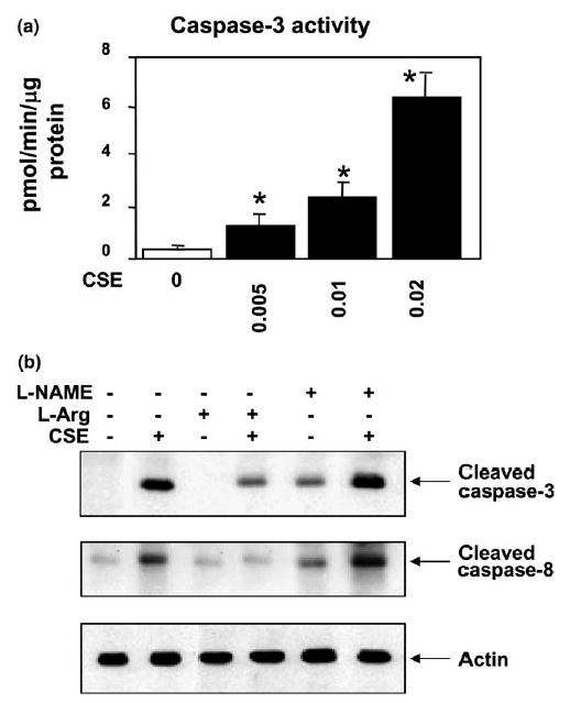
Fig. 3.

Effects of CSE on caspase-3 and -8 activations in HAECs. (a) Caspase-3 activity (means + S.E.M., N = 3) was increased with increasing CSE concentrations (F = 40.3, P = 0.01), and the caspase-3 activities treated with three different doses of CSE (0.005, 0.01 and 0.02 U) were significantly higher (*P < 0.01) than the control cells treated with standard medium only by the post hoc Dunnett’s test. (b) HAECs were treated with (lane 2) or without (lane 1) CSE (0.02 U) for 4 h. Treatment conditions are shown on the top of the figure. Cell lysates were separated on 12% SDS–PAGE. The membrane was immunoblotted with either anti-cleaved caspase-3 antibody (top panel of b) or anti-cleaved caspase-8 antibody (middle panel of b). CSE activated the cleavage of caspases-3 and 8 at 4 h. In pretreatment experiments, cells were incubated with 200 μM of l-arginine (lanes 3 and 4), or 400 μM of l-NAME (lanes 5 and 6) for 30 min followed by 0.02 U CSE exposure for 4 h (lanes 4 and 6). Pre-activation of eNOS by l-arginine attenuated the CSE-induced caspases-3 and 8 activations whereas inhibition of eNOS by l-NAME increased the CSE-induced caspase activation. Western blotting with anti-actin antibody was carried out to assess for equal loading (lower panel). Experiments were repeated three times and the representative electrophoresis is shown in the figure.
