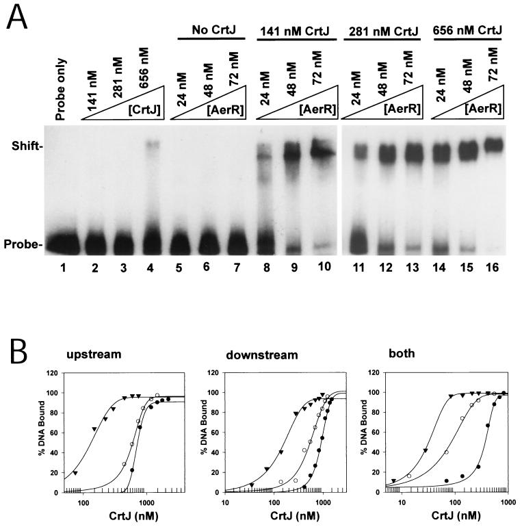
FIG. 6.
Cooperation between AerR and CrtJ in binding to the puc promoter region. (A) Gel mobility shift of CrtJ binding to the downstream puc palindrome in the absence or presence of AerR. Lane 1 is of probe only, while lanes 2 to 4 contain probe plus CrtJ at 141, 281, and 656 nM, respectively. Lanes 5 to 7 contain AerR at 24, 48, and 72 nM, respectively. Lanes 8 to 10 contain CrtJ at 141 nM in all lanes, as well as AerR at 24, 48, and 72 nM, respectively. Lanes 11 to 13 contain 281 nM CrtJ in each lane as well as AerR at 24, 48, and 72 nM, respectively. Lanes 14 to 16 contain 656 nM CrtJ in each lane as well as AerR at 24, 48, and 72 nM, respectively. (B) The graphs show a plot of the percentage of shifted 32P-labeled pucB probe versus the amount of CrtJ in the assay. Filled circles represent the DNA-binding isotherm of CrtJ with no AerR present, empty circles represent the CrtJ DNA-binding isotherm obtained in the presence of 24 nM AerR, and filled inverted triangles represent the CrtJ DNA-binding isotherm obtained in the presence of 48 nM AerR. The left, middle, and right graphs represent the binding curves obtained with a probe containing only the upstream CrtJ recognition palindrome, with a probe containing only the downstream recognition palindrome, and with a probe containing both recognition palindromes, respectively.

