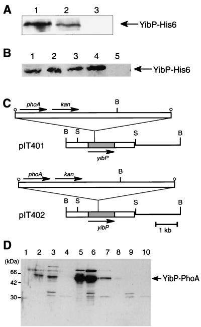
FIG. 6.
(A) Localization of the YibP-His6 fusion protein in cellular fractions. Cell extract of IT107 was fractionated into three fractions by the Sarkosyl method, and these fractions were analyzed by Western blotting using anti-His tag antibody. Each sample containing 10 μg of protein was applied to the gel. Lane 1, cytoplasmic fraction. Lane 2, inner membrane fraction. Lane 3, outer membrane fraction. (B) Effect of incubation with and without proteinase K on spheroplasts prepared from IT111 cells. Lane 1, total proteins of cells precipitated with trichloroacetic acid. Lane 2, spheroplasts. Lane 3, sonicated spheroplasts. Lane 4, spheroplasts incubated with proteinase K. Lane 5, sonicated spheroplasts incubated with proteinase K. (C) Insertion site of the TnphoA transposon in plasmids. B, BamHI; S, SacI. (D) C118 cells harboring pIT401 and pIT402 were exponentially grown in the absence of IPTG. Cell fractions prepared by the Sarkosyl method were analyzed by SDS-PAGE (15% gel) and Western blotting using anti-PhoA antibody. Lane 1, molecular size markers. Lanes 2 to 4, cellular fractions of cells with pIT401. Lanes 5 to 7, cellular fractions of cell with pIT402. Lanes 8 to 10, cellular fractions of cells without plasmid. Lanes 2, 5, and 8, cytoplasmic fraction. Lanes 3, 6, and 9, inner membrane fraction. Lanes 4, 7, and 10, outer membrane fraction.

