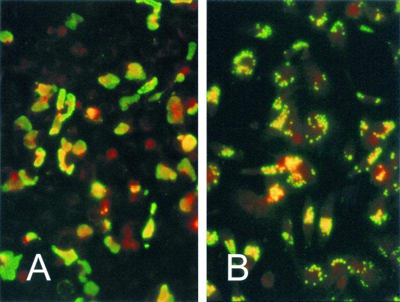
FIG. 1.
Immunofluorescence staining of Chp2-infected C. abortus (panel A) and CPAR39-infected C. pneumoniae (panel B). Monoclonal antibody 55 (which recognizes Chp2 VP1) was visualized with FITC-conjugated IgG. In C. pneumoniae, enlarged infected chlamydiae can be identified within inclusions; by contrast, in C. abortus, the whole inclusion is stained. Host cells are counterstained with Evans Blue, and both samples were taken at 72 h postinfection.

