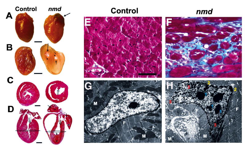
Figure 4.

(A) Gross appearance of a cardiomyopathic nmd heart (right) and an unaffected littermate control (left) (Scale, bar = 2 mm). (B) The same hearts sectioned longitudinally, showing the nmd heart (right) with grossly attenuated walls and dilated chambers containing one or more thrombi (↑ and * mark the limits of each thrombus) compared to the control heart (left). (C) Cross sections of hearts taken at the level depicted in panel D showing longitudinally sectioned hearts stained with H& E (Scale bar = 2mm). The nmd heart has one or more organizing mural thrombi (⇒ and * mark the limits of each thrombus). Also, note the attenuated papillary muscles (⇑ in C and D) in the nmd versus control heart. Masson’s Trichrome stained histological samples of the ventricular myocardium (F) from an nmd mouse displayed severe interstitial fibrosis, indicated by the blue stained collagen, compared to the control in panel E (Scale, bar = 25 μm). Panel G shows an electron micrograph of a normal cardiomyocyte nucleus, while panel H shows an nmd ventricular cardiomyocyte nucleus. Morphological alterations include multiple nuclear condensations in addition to the nucleolus (1), chromatin margination (2), and marked ruffling of the nuclear membrane (3) suggestive of an apoptotic process. (M = mitochondria, F = myofibrils; Magnification = 6,700x).
