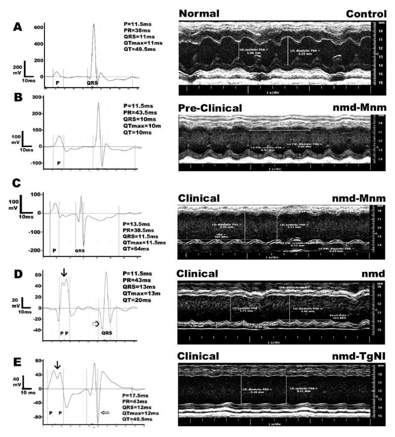
Figure 6.

Representative electrocardiograms and M-mode echocardiographic images in 5–7 week old nmd mice with different clinical presentations based on normal/elevated levels of creatine kinase cardiac isoenzyme CK-MB. Panel A represents a normal B6 control. In panels B and C, the same modified nmd-MnmC mouse was tested before clinical symptoms were evident at 5 wks of age and again following a significant rise in its CK-MB values at 7 wks ( Table 1). Panels D and E were obtained from an nmd or transgenic nmd-TgNI mouse with overt clinical signs, respectively. The three mice with clinical heart failure showed similar changes in echocardiographic indices compared to the control mouse ( Table 1 and A, C, D and E). In contrast, the mouse in panel B, despite an apparently normal CK-MB level (Table 1), showed intermediate echocardiographic indices. The representative electrocardiograms further validate the presence of cardiomyopathy and its progression toward heart failure. Note also the various scales in each electrocardiogram (mV), which were necessary in order to show each individual tracing in greater detail.
