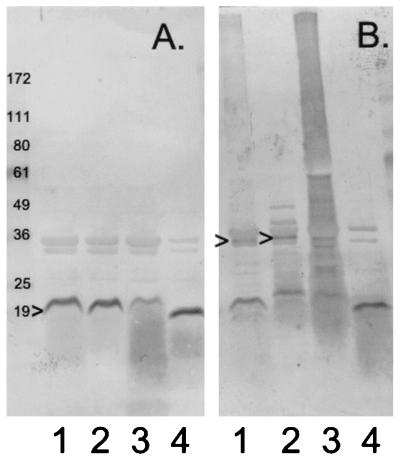
FIG. 2.
Accumulation of TraV proteins in the outer membrane. (A) Standard (reducing) gel; (B) nonreducing gel. Outer membrane fractions were obtained by banding in sucrose density gradients as described previously (9). Gels contained 4 to 20% polyacrylamide gradients. For nonreducing gels, mercaptoethanol was omitted from the sample buffer. Lanes 1, outer membrane from pOX38 traV::cat/pUC traV (wild-type) cells; lanes 2, outer membrane from pOX38 traV::cat/pUC traVC10S cells; lanes 3, outer membrane from pOX38 traV::cat/pUC traVC18S cells; lanes 4, outer membrane from pOX38 traV::cat/pUC traVC10S/C18S cells. The caret at the left of panel A indicates the TraV protein. The carets in panel B indicate possible TraV dimers. Numbers to the left of the figure are molecular masses in kilodaltons of marker proteins with the indicated mobilities.

