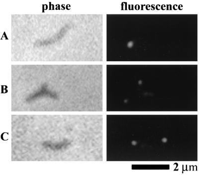
FIG. 8.
Localization of P65 protein in wild-type M. pneumoniae (A), mutant I-2 (B), and an hmw3::Tn4001 transformant (C). On the left are individual cells viewed by phase-contrast microscopy, and on the right are the corresponding fluorescent images. The contrast was adjusted to compensate for protein level differences among the strains (see Fig. 3E).

