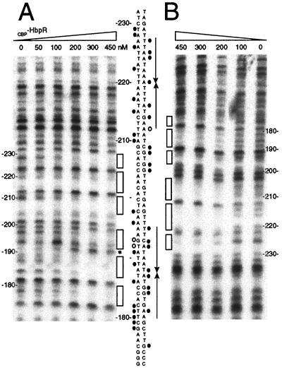
FIG. 4.
DNase I footprinting analysis of CBP-HbpR binding to UASs C-1 and C-2. The 229-bp 32P-end-labeled fragment hbpCCC6 containing UASs C-1 and C-2 was incubated with increasing amounts of CBP-HbpR (0 to 450 nM). (A) CBP-HbpR-mediated DNase I protection pattern for the bottom strand. (B) CBP-HbpR-mediated DNase I protection pattern for the top strand. Boxes indicate the regions protected from DNase I digestion upon the addition of CBP-HbpR. Between the panels, the sequences of UASs C-1 and C-2 and the positions that were contacted by CBP-HbpR are shown. Black circles indicate protection from DNase I digestion, while open circles and the asterisk indicate increased sensitivity to DNase I. The positions of the palindromes are indicated by arrows. Nucleotide numbering was relative to the transcriptional start site of hbpC.

