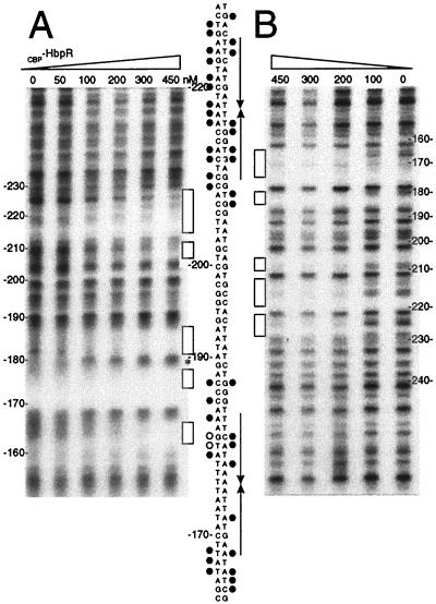
FIG. 5.
DNase I footprinting analysis of CBP-HbpR binding to UASs D-1 and D-2. The 256-bp 32P-end-labeled fragment hbpD3D4 containing UASs D-1 and D-2 was incubated with increasing amounts of CBP-HbpR (0 to 450 nM). (A) CBP-HbpR-mediated DNase I protection pattern for the bottom strand of hbpD3D4. (B) CBP-HbpR-mediated DNase I protection pattern for the top strand of hbpD3D4. Nucleotide numbering was relative to the hbpD transcriptional start site. For symbols and further explanations, see the legend to Fig. 4.

