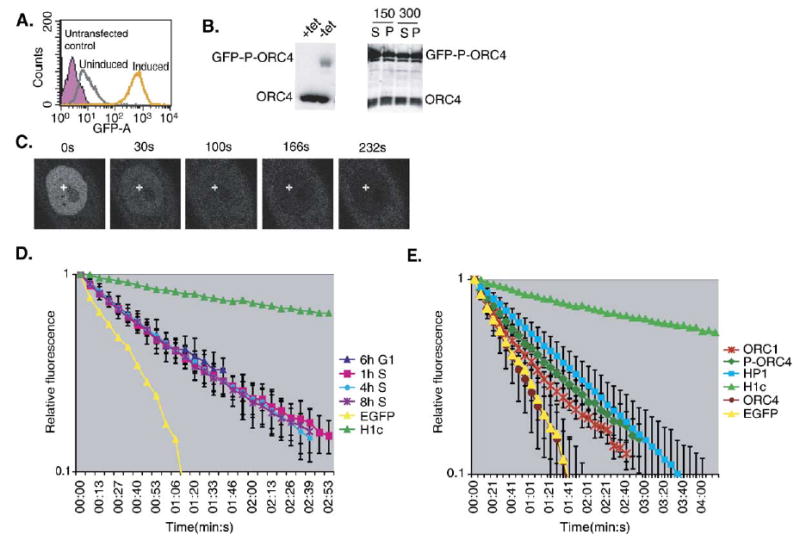
Fig. 5.

Cell line characterization and FLIP of ORC4. (A) Homogeneity and inducibility of GFP-P-ORC4. PC1–11 cells (CHO cells stably expressing GFP-P-ORC4) were grown either in the presence (uninduced—gray) or absence (induced—orange) of tetracycline and analyzed for EGFP expression by FACS. Untransfected CHO cells (purple) were used as a negative control. (B) The expression of GFP-P-ORC4 is inducible (left panel) and GFP-P-ORC4 exhibits similar salt extractability as endogenous ORC4 (right panel). Whole cell extracts from induced and uninduced PC1-11 cells were run on 10% SDS-PAGE gels and immunoblotted with anti-ORC4 demonstrating induction of full-length GFP-P-ORC4 (left panel). PC1-11 cells were induced and isolated nuclei fractionated using increasing levels of NaCl (150 mM and 300 mM) and the resulting fractions were probed with anti-ORC4 antibodies (right panel). (C) Typical images from the cell-cycle FLIP of GFP-P-ORC4 in PC1-11 as in Figs. 4A and C. (D) FLIP kinetic plot from the cell-cycle FLIP analysis of GFP-P-ORC4, as described in Fig. 4. The error bars represent the standard deviation from one experiment. For each experiment, at least 5 cells were photobleached with 2–3 measurements taken per cell. EGFP and histone H1c are provided as reference marks for mobility. Similar results were obtained in three independent experiments. (E) Comparative summary of the relative FLIP kinetics for GFP-ORC1 (ORC1), GFP-P-ORC4 (P-ORC4), ORC4 (without the peptide linker) and HsHP1α.
