
Fig. 1.
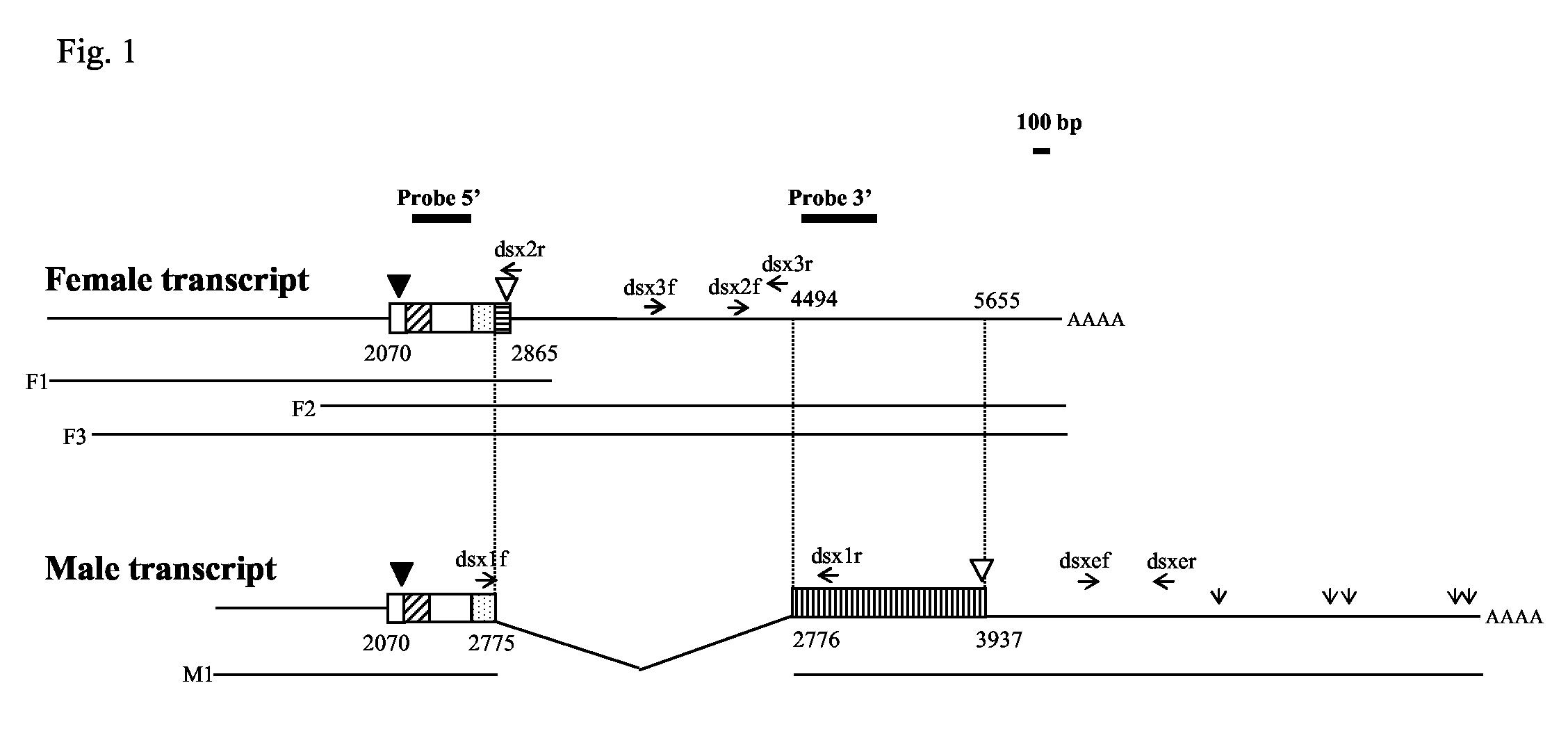
Schematic representation of the putative female (F1, F2 and F3) and male (M1) Agdsx transcripts isolated from a mixed pupal A. gambiae cDNA library. The sequence of the putative female transcript was deduced by combining the information provided by clones F1, F2 and F3, while the sequence of the putative male transcript was based on clone M1. The diagonally hatched box represents the DBD/OD1 domain, and the stippled box represents the non-sex-specific region of the OD2 domain. The putative female- and male-specific regions are indicated by a horizontal and a vertical hatched box, respectively. Black and white triangles indicate the position of the initiation and stop codon, respectively. Open vertical arrows in the male transcript indicate polyadenylation signal sequences. Numbers indicate the distance in bp from the start of clone F1, which was arbitrarily considered as the start for both female and male transcripts. The location of Probe 5’ and Probe 3’ is indicated by solid lines. Primers used in RT-PCR experiments are indicated as horizontal arrows. The bar (100 bp) indicates the scale of the figure.