
Figure 3.
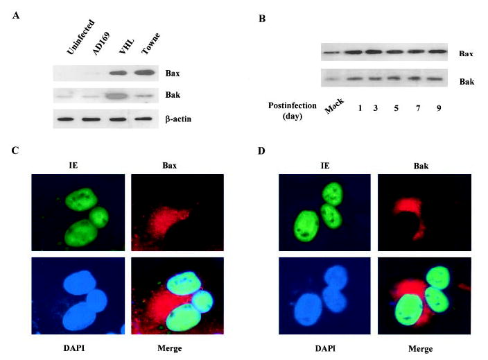
Increased Bax and Bak levels in HCMV-infected endothelial cells. A, Increase of Bax and Bak expression in HCMV-infected endothelial cells. Total protein from uninfected (cultured for the same duration) and infected (by AD169, VHL/E, and Towne strains) HAECs (day 9) was extracted and blotted with anti-Bax and anti-Bak antibodies. B, Time-dependent increase of Bax and Bak expression in HCMV-infected cells. After indicated infection time, total protein from mock-infected and VHL/E-infected HAECs was extracted and blotted with anti-Bax and anti-Bak antibodies. C, Increased Bax level in HCMV-infected HAECs. VHL/E-infected HAECs (day 9) were stained with anti-IE antibody-FITC (green), anti-Bax antibody-Texas Red (red), and DAPI (blue). Merged image shows colocalization. D, Increased Bak level in HCMV-infected HAEC. VHL/E-infected HAECs (day 9) were stained with anti-IE antibody-FITC (green), anti-Bak antibody-Texas Red (red), and DAPI (blue). Merged image shows colocalization.