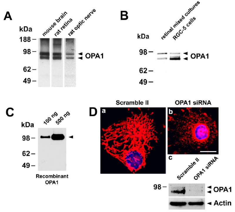
Fig. 1.

A: OPA1 expression in the normal retina and optic nerve. Immunoblot of extracts from mouse brain, rat retina, and optic nerve probed with OPA1 antibody. B: OPA1 expression in the normal retinal mixed cultures and RGC-5 cells shown by immunoblotting. The arrows show the positions, based on comparison with size standards, of the 90 and 80 kDa forms of OPA1. C: Immunoblotting of recombinant human OPA1 protein using OPA1 antibody. D: HeLa cells transfected with scramble II, control siRNA (a), or with OPA1 siRNA (b). Mitochondrial morphology and nuclei were identified by colabeling with MitoTracker Red and Hoechst. (c) OPA1 immunoblot of extracts from HeLa cells transfected with scramble II siRNA or OPA1 siRNA. The blot was stripped and reprobed with anti-actin antibody to ensure similar protein loading. Scale bar = 10 μm in a (applies to a,b).
