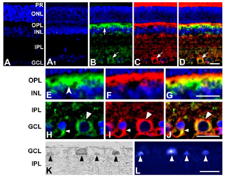
Fig. 2.

Cellular localization of OPA1 in the normal rat retina. A: Control. When the primary antibody was omitted, there was no binding of the secondary antibody. A1: Immunohistochemistry of rat retina using OPA1 antibody preabsorbed with OPA1 peptide. No OPA1 immunoreactivity was detected. B–G: OPA1 (B,E; green) and cytochrome c (C,F; red) double immunohistochemistry. OPA1 immunoreactivity (OPA1-IR) was seen in the OPL, INL, IPL, and GCL. Note that neurons were positive for OPA1 in the OPL (concave arrow) and GCL (arrow). Cytochrome c, a marker for the mitochondrial intermembrane space, colocalized with OPA1 (D). Higher magnification showed that OPA1-IR is present in horizontal cells (concave arrowhead) in the OPL (E) and OPA1-IR colocalized with cytochrome c-IR in cells with large (large arrowhead) or small (small arrowhead) sized soma (J). K,L: OPA1 (K, HRP-DAB) and Fluoro-Gold (L) double labeling. Neurons containing OPA1-IR were colabeled by Fluoro-Gold (arrowheads), indicating that retinal ganglion cells in the GCL contained OPA1 protein. HRP-DAB, horseradish peroxidase-diaminobenzidine; IR, immunoreactivity; PR, photoreceptor; ONL, outer nuclear layer; OPL, outer plexiform layer; INL, inner nuclear layer; IPL, inner plexiform layer; GCL, ganglion cell layer. Scale bars = 20 μm in D (applies to A–D), G (applies to E–G), J (applies to H–J), L (applies to K,L).
