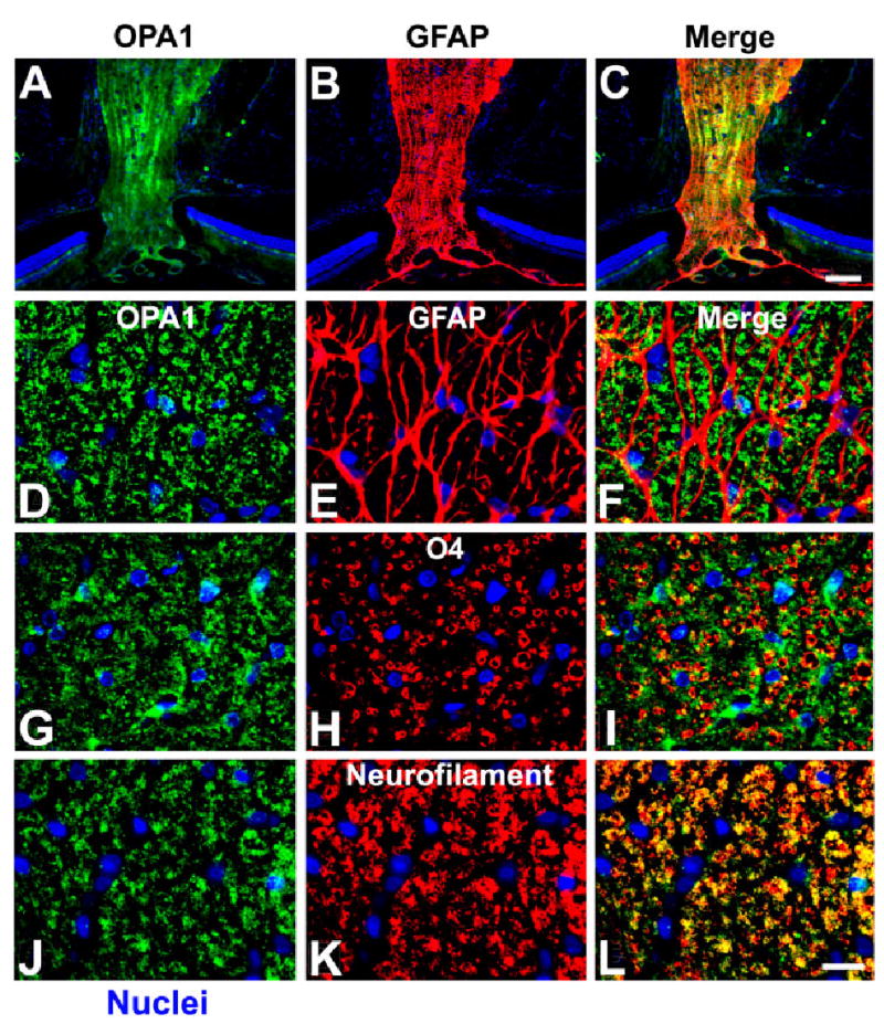
Fig. 4.

Cellular localization of OPA1 in the normal rat optic nerve. A–F: OPA1 (A,D) and GFAP (B,E) double immunohistochemistry. Cells positive for OPA1 were not positive for GFAP, an astrocyte marker (F). G–I: OPA1 (G) and O4 (H) double immunohistochemistry. OPA1 cells were not positive for O4, an oligodendrocyte marker (I). J–L: OPA1 (J) and neurofilament (K) double immunohistochemistry. OPA1-positive cells were also positive for neurofilament, indicating that OPA1 was present in axons of the optic nerve (L). Scale bars = 100 μm in C (applies to A–C); 20 μm in L (applies to D–L).
