
Figure 4. The mechanisms of CD8+ T-cell tolerance induction by ImC.
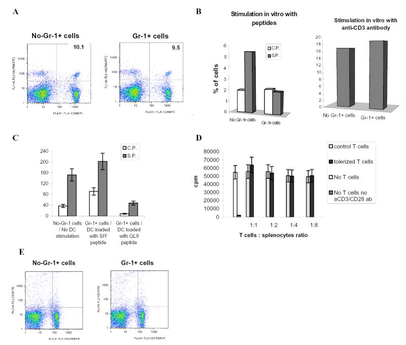
A. Adoptive transfer of OT-1 cells, Gr-1+ cells and immunization with peptide was done as shown above in Fig.1A. LN cells were isolated on day 10 after immunization and percentage of antigen-specific CD8+Valpha2+ T cells was determined by flow cytometry. A representative experiment of three performed is shown.
B. Adoptive transfer of OT-1 cells, Gr-1+ cells and immunization with peptide was done as shown in Fig.1A. Draining LNs were re-stimulated in vitro with specific (S.P.) or control (C.P.) peptides (10 μg/ml) or anti-CD3 antibody (1μg/ml) for 48 h. Production of IFN-γ was measured using intracellular staining within antigen-specific CD8+Vα2+ T cells. Typical results of one of two performed experiments are shown.
C. 2C T cells (3 x 10⁶) were adoptively transferred i.v. into naive C57BL/6 recipients followed by 3 days later immunization with 2C specific peptide SIYRYYGL (100 μg) and injection of Gr-1+ cells (3 x 10⁶) from EL-4 tumor-bearing mice. DCs generated in vitro from control C57BL/6 mice were activated with LPS, pulsed with either specific (SIY) or control (QL9) peptides, and injected s.c. (4x10⁵) into mice 10 days after first immunization. In 7 days after that mice were sacrificed, LN cells isolated and re-stimulated with specific (S.P.) or control (C.P.) peptides. The number of IFN-γ producing cells was measured by ELISPOT. Results presented as Mean±SD.
D. T cells isolated from OT-1 mice were transferred i.v. into naïve C57BL/6 mice. Mice were immunized with SIINFEKL together with the transfer of Gr-1+ isolated from spleens of EL-4 tumor-bearing mice. Ten days later LN cells were collected and T cells were isolated and added in triplicates at indicated ratio to 105 splenocytes isolated from naïve mice and incubated for 4 days with 0.5 μg/ml anti-CD3 antibody and 0.1 μg/ml anti-CD28 antibody. Cell proliferation was evaluated using 3H-thymidine uptake. Results presented as Mean±SD. Control T cells – T cells isolated from immunized mice without transfer of Gr-1+ImC, tolerized T cells – T cells isolated from mice after adoptive transfer of Gr-1+ cells.
E. Adoptive transfer of OT-1 cells, Gr-1+ cells and immunization with peptide was done as shown in Fig.1A. LN cells were isolated on day 9 after immunization and percentage of CD4+CD25+ cells was determined by flow cytometry. Typical results of one of two performed experiments are shown.