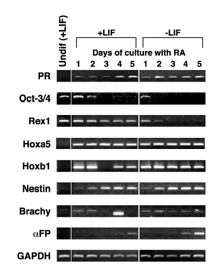
Figure 2.

RA induction of PR expression in CCE mES cells. RNA was prepared from cells cultured as described in Materials and Methods and RT-PCR performed using primers and conditions listed in Table I. Samples from mES cells maintained in the undifferentiated state by addition of LIF to the culture media are labeled Undif (+LIF). Differentiation was induced by RA for the time indicated in plated cells grown in the presence or absence of LIF. The expression of a series of mES cell differentiation markers is also shown. Note that the band on day 4 of +LIF in the Brachy panel is smaller than the correct product and represents a primer dimer. GAPDH is used as a normalization control.
