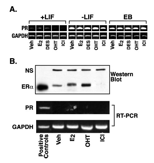
Figure 5.

Progesterone receptor expression in mES cells is regulated by differentiation, rather than estrogen receptor signaling. A. RNA was prepared from cells cultured as described in Materials and Methods and RT-PCR performed using primers and conditions listed in Table I. Samples from mES cells were maintained in the undifferentiated state by addition of LIF to the culture media (+LIF). Differentiation was induced by LIF withdrawal for the 4 days in plated cells (−LIF) or 8 days in EBs (EB). Hormone treatments were vehicle control (veh), 5 nM E2, 3.7 μM DES, 100 nM OHT, or 100 nM ICI for 4 days in plated cells or the final 2 days in EBs. B. Undifferentiated mES cells were transfected with an hERα expression vector for 24 hrs as described in Materials and Methods. Cells were treated with hormone as in (A) for 22 hrs and replicate cultures harvested for protein or RNA. Western blot analysis for ERα was performed as described in Materials and Methods. The band labeled NS (non-specific) acts as a loading control for the mES cell extract lanes. The positive control is 10 fmoles purified hERα from PanVera. The positive control for PR expression in the RT-PCR panel is RNA from mES cells withdrawn from LIF for 4 days.
