Figure 6.
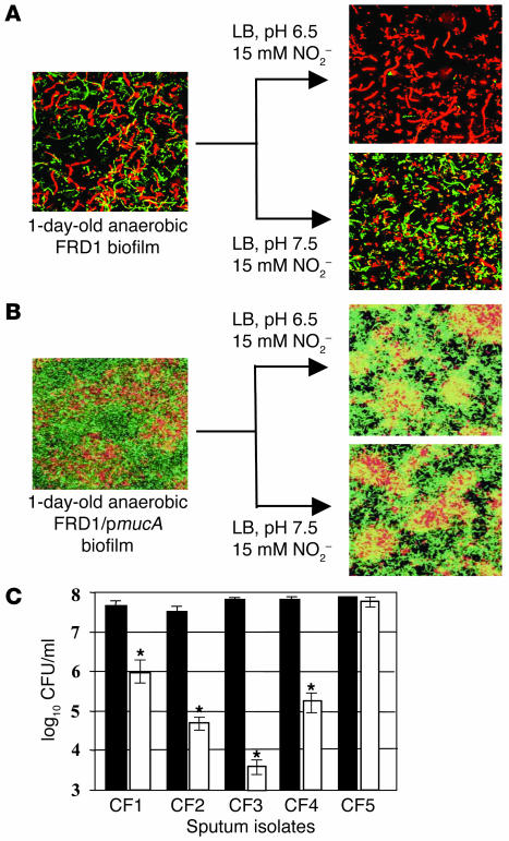
Effect of HNO2 on killing P. aeruginosa in biofilms and fresh sputum isolates. (A) Confocal laser microscopic analysis of anaerobic FRD1 biofilms. Live cells are stained with syto-9 (green), and dead cells are stained with propidium iodide (red). Top (x-y plane) views are projected from a stack of 125 images taken at 0.4-μm intervals for a total of 50 μm. Before staining, 1-day-old anaerobic FRD1 biofilms were treated anaerobically with 15 mM NO2– at pH 6.5 and 7.5 for 2 days. (B) Biofilms were grown as in A using FRD1/pmucA. (C) Toxicity of 15 mM NO2– (pH 6.5) toward P. aeruginosa sputum isolates. Viable cells in the initial inoculum (black bars) and after a 24-hour anaerobic incubation (white bars) are presented in logarithmic scale. CF1–CF4 are mucoid mucA mutants, and CF5 is nonmuocid and possesses a wild-type mucA gene. Each strain was tested in triplicate and the mean ± SEM is presented. *P < 0.05.

