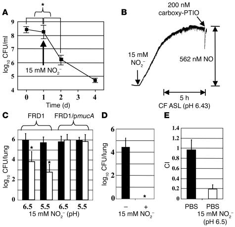
Figure 7.
Applications of HNO2-mediated killing of mucoid P. aeruginosa to clinical specimens. (A) Killing of FRD1 by NO2– in sterile ultrasupernatants of CF airway secretions derived from explanted CF lungs. Bacteria were incubated anaerobically for 24 hours, and 15 mM NO2– was added. CFUs were determined (n = 3) and plotted as the X ± SEM versus time. *P < 0.01 versus CFU decrease before adding NO2–. (B) NO generation in CF ASL by 15 mM NO2–. Except for the media and pH, experimental conditions were identical to those used in Figure 4B. (C) Effects of NO2– on killing of FRD1 and FRD1/pmucA in mouse lungs. CD1 mice were infected with FRD1 or FRD1/pmucA as described in Methods. Infected mice were treated with buffer (black bars) and buffered NO2– (white bars) daily, and viable bacteria from the lung homogenates were enumerated. n = 8 per group. *P < 0.01 versus buffer alone. (D) Effects of long-term NO2– treatment on killing of FRD1 in mouse lungs. Another group of FRD1-infected mice were treated daily with buffer (–, 50 mM sodium phosphate, pH 6.5) or buffer with 15 mM NO2– (+) for 16 days (n = 8 per group). Organisms surviving treatment with buffer and NO2– were shown in logarithmic scale. *P < 0.01 versus buffer alone. (E) CI experiments with 106 FRD1 and FRD1/pmucA intratracheally instilled into CD1 mouse airways and incubated for 6 days prior to harvesting of mouse lungs and enumeration of CFUs after homogenization.

