
Fig. 6. Effect of pharmacological inhibitors on trans-10, cis-12 CLA-mediated activation and nuclear accumulation of P-ERK1/2.
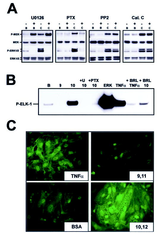
Cultures of SV cells containing newly differentiated human adipocytes were serum starved for 24 h and then treated as follows. A, cultures were pretreated with the MEK inhibitor U0126 (10 μm), the GPCR-Gi/o coupling inhibitor PTX (100 ng/ml), the c-SRC kinase inhibitor PP2 (1 μm), or the protein kinase C inhibitor calphostin C (Cal. C, 200 nm) for 1 h and subsequently treated with either a BSA vehicle (B) or 30 μm trans-10, cis-12 CLA complexed to BSA (C) for an additional 24 h. Cell extracts were immunoblotted for the active phosphorylated forms of MEK (P-MEK) and ERK (P-ERK1/2) and subsequently were stripped and reprobed with antibodies recognizing total MEK (MEK), and total ERK (ERK1/2). Data shown are representative of two to three independent experiments for each panel. B, cultures were pretreated with a vehicle (dimethyl sulfoxide) for 1 h, 10 μm U0126 for 1 h, 100 ng/ml PTX for 1 h, or 1 μm BRL49653 for 24 h, and subsequently treated with a BSA vehicle (B), 30 μm cis-9, trans-11 CLA complexed to BSA (9), or 30 μm trans-10, cis-12 CLA complexed to BSA (10) for an additional 24 h. Active ERK was then immunoprecipitated from total cell extracts and used in an in vitro kinase reaction with its known substrate, recombinant ELK-1. The resulting kinase reaction was subjected to SDS-PAGE and probed for phosphorylated ELK-1 using a phospho-specific antibody. A 30-min TNF-α treatment (100 ng/ml) of human adipocytes and active ERK-2 (ERK) was used as positive control for enzyme activation. C, cultures were pretreated with either a BSA vehicle control, 30 μm cis-9, trans-11 CLA (9,11), or 30 μm trans-10, cis-12 CLA (10,12) for 24 h. A 30-min treatment with TNF-α was used as a positive control for MAPK activation. Active ERK1/2 was then detected using immunofluorescence microscropy. Data shown are representative of two to three independent experiments for each panel.