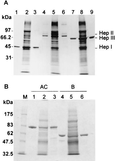
FIG. 5.
Coomassie blue R-250 analysis of HepI, -II, and -III and ChnA and -B purified from either the native or transconjugant F. heparinum strain. (A) SDS-10% PAGE Coomassie blue R-250-stained gel. Lane 1, 1 μg of purified native HepI; lane 4, native HepII; and lane 7, native HepIII. Lane 2, 0.1 OD600 units of soluble FIBX5; lane 5, same amount of FIBX3; and lane 8, FIBX4 cell extract. Lane 3, 1 μg of purified tHepI; lane 6, same amount of tHepII; and lane 9, same amount of tHepIII. (B) SDS-7.5% PAGE Coomassie blue R-250-stained gel. Lane M, molecular weight marker; lane 1, 1 μg of purified native ChnA; lane 4, 1 μg of native ChnB; lane 2, 0.1 OD600 units of soluble FIBX6; lane 5, same amount of FIBX7; lane 3, 1 μg of purified tChnA; and lane 6, same amount of tChnB.

