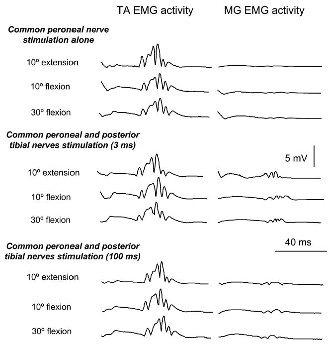
Fig. 3.

Full-wave rectified EMG activity in TA and MG muscles is shown for one subject (S3) under all conditions of testing. EMG activity of these muscles is shown with hip positioned at different angles of flexion and at 10° of extension during common peroneal nerve (CPN) stimulation alone and following conditioning of the soleus H-reflex (posterior tibial nerve stimulation) with CPN stimulation at 3 ms and at 100 ms of conditioning test intervals. Under all conditions motor axons were not excited while TA displayed a stable in amplitude H-reflex in all hip angles investigated
