Fig. 2.

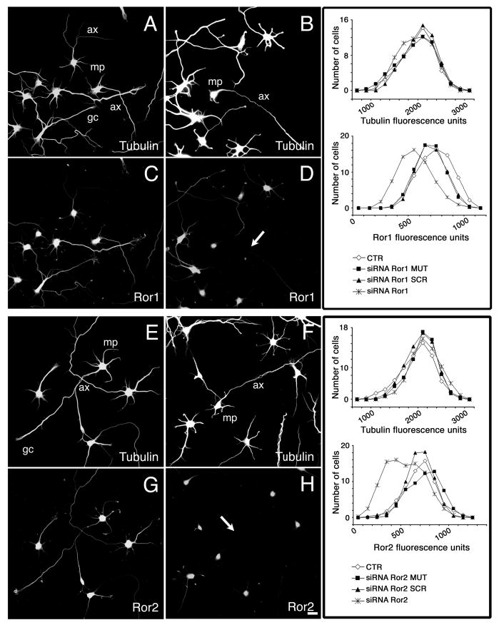
Ror1 and Ror2 suppression by RNAi in cultured hippocampal neurons. (A-H) 2- day cultured control (A,C,E,G), siRNA Ror1- (B,D) and siRNA Ror2- (F,H) treated hippocampal neurons were double stained with tubulin (A,B,E,F) and Ror1 (C,D) or Ror2 (G,H) antibodies. Transfection with the cognate siRNA duplex reduced Ror1 and Ror2 expression in neurons from the targeted cultures (arrows in D,H), leaving their tubulin expression unaffected (B,F). Panels on the right represent the distribution function of the immunoreactivity pixel intensity of Ror1 and tubulin (upper panel) and Ror2 and tubulin (lower panel) in control and targeted hippocampal neurons. Controls included non-transfected neurons (CTR) and neurons transfected with oligomers carrying a 2 base pair mutation (siRNA MUT) or scrambled oligomers (siRNA SCR). Note the shift in the expression levels of Ror1 and Ror2, but not of tubulin, in the targeted cultures when compared to controls. ax, axon; gc, growth cone; mp, minor process. Bar, 20 μm.
