
Fig. 5. 1H-13C HSQC spectrum of Gα protein labeled at Leu-348, Gly-352, and Phe-354.
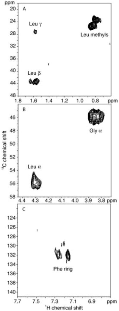
The panels display the regions containing the Leu-348 methyl Cβ-H and Cγ-H resonances. The Gly-352 (A) and Leu-348 (B) Cα-H resonances and the Phe354-13C-ring resonances (C).