
Figure 3.
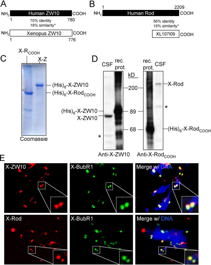
Characterization of Xenopus ZW10 and Rod. (A and B) Schematic alignment of Xenopus and human ZW10 (A) or Xenopus RodCOOH (XL107l09) and human Rod (B). Amino acid positions as well as percentage identity and additional (*) similarity on the protein level are indicated. (C) Coomassie staining of purified recombinant X-ZW10 (X-Z) and X-RodCOOH (X-RCOOH). (His)6-tagged proteins were purified from insect cells and analyzed by Coomassie blue staining. (D) Immunoblot analysis of pAbs to X-ZW10 and X-RodCOOH. 20 ng of recombinant protein (rec. prot.) and 1 μl CSF extract were analyzed by immunoblot with affinity purified anti–X-ZW10 (1348) or anti–X-RodCOOH (1351). Position of the endogenous frog proteins in the CSF extract is indicated. Cross-reacting proteins are marked by asterisks. (E) Immunolocalization of X-ZW10 and X-Rod. Sperm nuclei replicated in cycled CSF extract were immunostained for X-BubR1 and X-ZW10 or X-Rod. DNA (DAPI) is in blue. Enlarged boxes show overlap of X-BubR1 and X-ZW10–X-Rod signals on a sister kinetochore pair.