
Figure 3.
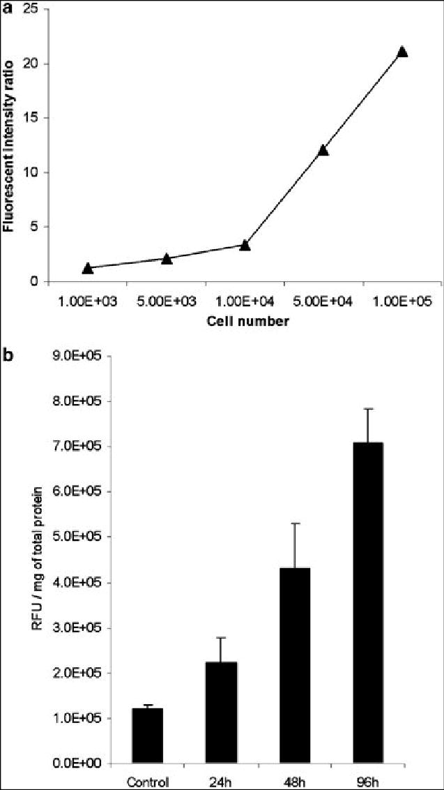
Fluorimetry. The fluorescent signal from NV1066 infected cells increased linearly with cell number in vitro (A), suggesting that fluorescent intensity could be used as a surrogate for infected cell number. Fluorescent intensity ratios (fluorescence of infected cells/fluorescence of uninfected cells) reach sufficient levels to visually distinguish induced fluorescence from background fluorescence with as few as 1 x 10⁴ cells infected with NV1066 in vitro. In ex vivo experiments, fluorescence increased over time in tumors infected with NV1066, representing viral replication and spread within the tumor (B). By 48 hours after infection, significant differences in the fluorescent signal were present between control and infected tumors (p = .04)