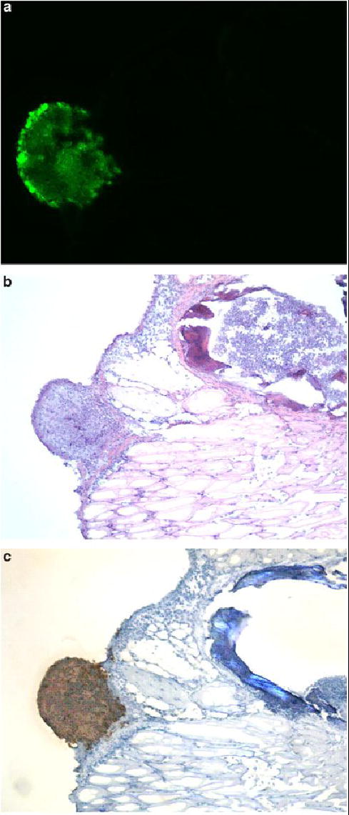
Figure 7.

Viral specificity for tumor. Tissue specimens were selected by EGFP expression under fluorescent stereomicroscopy in intact animals. Serial sectioning was performed and specimens were examined under fluorescent microscopy (A), then H & E stained (B) for identification of tumor cells. All sections that expressed EGFP had tumor cell infiltrates. Staining for polyclonal HSV-1 antibody corresponded to areas of EGFP expression and tumor (C). No viral staining was evident in tissues that did not express EGFP.
