
Figure 1.
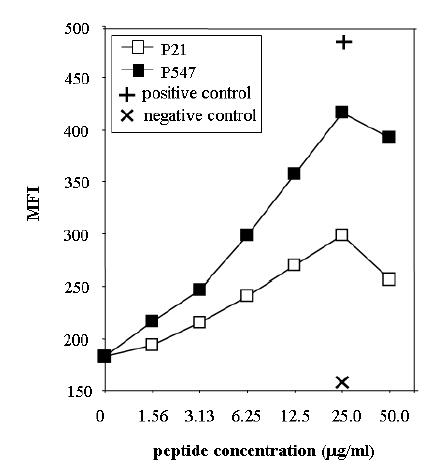
Binding of mesothelin peptides to HLA-A2. Peptides were analyzed for binding to T2 cell line as described in “Materials and Methods.” Peptides were used at concentrations of 0 to 50 μg/ml. P21 peptide (open square), P547 (solid square), positive control (MUC-1 peptide) (+) and negative control (HLA-A3 binding peptide) (X). Results are expressed in mean fluorescence intensity (MFI).