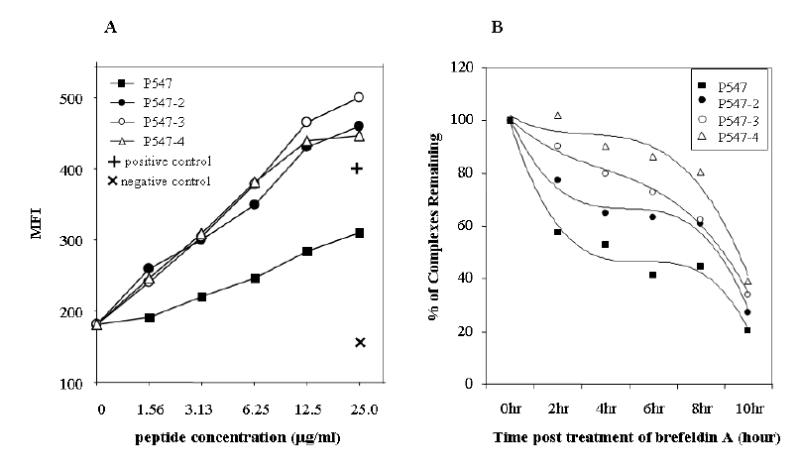
Figure 2.

Binding of a native mesothelin peptide and its agonist peptides to HLA-A2. Peptides were analyzed for binding to T2 cell line as described in “Materials and Methods.” (A) Peptides were used at concentrations of 0 to 50 μg/ml. P547 peptide (solid square), P547-2 (solid circle), P547-3 (open circle) and P547-4 (open triangle), positive control (MUC-1 peptide) (+) and negative control (HLA-A3 binding peptide) (X). Results are expressed in mean fluorescence intensity (MFI). (B) Comparison of the stability of HLA-A2 peptide complexes of mesothelin native and agonist peptides. T2 cells were incubated overnight with P547 (solid square), P547-2 (solid circle), P547-3 (open circle), and P547-4 (open triangle) peptides at a concentration of 25 μg/ml and then were washed free of unbound peptide and incubated with brefeldin A to block delivery of new class I molecules to the cell surface. At the indicated times, cells were stained for the presence of surface peptide-HLA-A2 complexes. Results are expressed in relative percentage of binding compared with 100% at time 0.
