
Fig. 5.
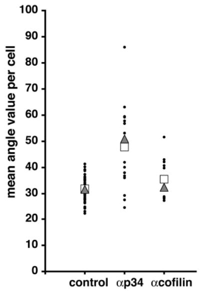
The filament incidence angles at the membrane are altered in anti-p34 injected cells. The value of the incidence angle of free end filaments at the leading edge 1 minute after stimulation was measured. Values plotted are mean angle value per cell (filled circle), median (triangle) and mean (square). (See Table 1 for statistics.)