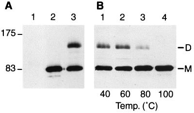
FIG. 1.
PBP1a dimer and stability. (A) Membrane fractions prepared from strain QCA2 (25 μg, lane 1) or strain QCA2(pPONAMyc) (25 μg, lanes 2 and 3) were incubated for 10 min in sample buffer at 100°C (lanes 1 and 2) or room temperature (lane 3), submitted to SDS-PAGE, and analyzed by Western blotting with the anti-c-myc 9E10 monoclonal antibody. The molecular mass markers (in kilodaltons) are displayed on the left. (B) Membrane fractions (25 μg) prepared from strain QCA2(pPONAMyc) were incubated for 10 min in sample buffer at temperatures ranging from 40 to 100°C as indicated, submitted to SDS-PAGE, and analyzed by Western blotting with the anti-c-myc 9E10 monoclonal antibody. M, monomeric form of PBP1a; D, dimeric form of PBP1a.

