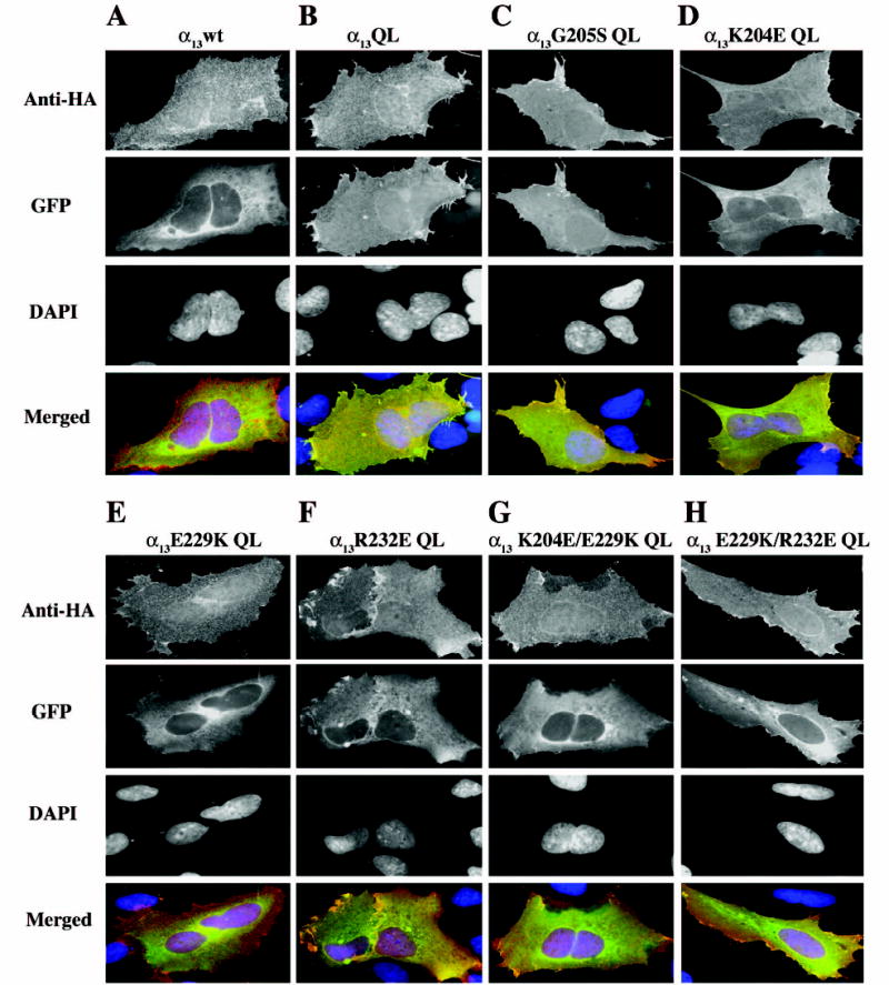
Fig. 5. Subcellular localization of p115RhoGEF in cells co-expressing constitutively active α13 mutants.

HEK293 cells were transiently co-transfected with 0.5μg each expression plasmid for the indicated α13 constructs and 0.1 μg DNA encoding p115RhoGEF-GFP. 48 h after transfection, cells were fixed and stained with a monoclonal anti-HA antibody, followed by Alexa-594-conjugated anti-mouse antibody, to stain for the HA epitope tagged α13 constructs (anti-HA panels). p115RhoGEF’s localization was determined by visualization of the intrinsic fluorescence of GFP (GFP panels), and DAPI staining was used to identify nuclei (DAPI panels). Three color merged images show α13 (red), p115RhoGEF-GFP (green) and nuclei (blue) (Merged panels). A representative image of more than 100 cells analyzed in three separate experiments is shown.
