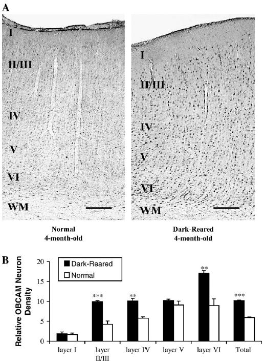
Figure 10.

(A) Photomicrographs illustrating the effect of dark-rearing on OBCAM-immunoreactivity in the primary visual cortex of a normal and a dark-reared cat at 4 months of age. More OBCAM-immunopositive neurons were present in the primary visual cortex of the dark-reared animal. (B) Histograms that show the number of OBCAM-immunopositive neurons in visual cortex of 4-month-old normal and dark-reared animals. Black columns: dark-reared; white columns: normal. Other conventions are the same as in Figure 8. **P < 0.01, ***P < 0.001 (ANOVA analysis).
