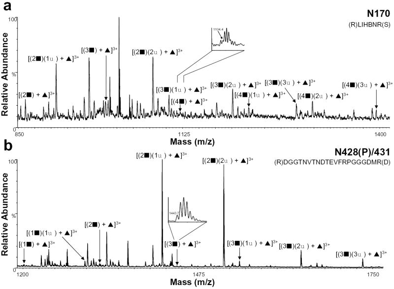
Figure 2.
Positive ion nHPLC/ESI/MS spectra summed over the chromatographic retention window in which complex glycopeptides a) N170 and b) N428(P)/431 eluted. The inset in panel a is for the ion corresponding to [(4▪) + ▴]3+. The inset in panel b is for the ion corresponding to [(3▪) + ▴]3+. Both insets are of spectra summed over the elution window of that component. For figure clarity, the peaks corresponding to other tryptic peptides/glycopeptides are not labeled. ▪ = GlcNAc-Gal, ♦ = sialic acid, and ▴ = fucose; the number before each symbol indicates how many of each are present. The superscripted number is the charge state. In the amino acid sequence B= carboxymethylcysteine.

