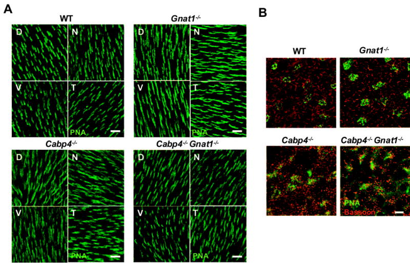
Figure 3.

PNA labeling of cones in retinal whole-mounts of 2-month-old mice. (A) Staining of cone outer segments in the four quadrants of flat-mounted retina labeled with PNA from wild-type and all knockout mice. D, dorsal; V, ventral; N, nasal, T, temporal. Scale bars, 20 μm. (B) Double labeling with anti-bassoon (red) and PNA (green) across the outer plexiform layer in the temporal quadrant of the flat-mounted retina. Horseshoe-shaped structures are observed in wild-type and Gnat1−/− mice, but mostly punctate staining is observed in Cabp4−/− and Cabp4−/−Gnat1−/− mice. In wild-type and Gnat1−/− mice, the PNA-labeled cone pedicles are round, but the cone pedicles of Cabp4−/− and Cabp4−/− Gnat1−/−mice are more disorganized and appear spread out in a diamond-shaped structure. Scale bar, 5 μm.
