
Fig. 7. AFM of native discs and disc membranes isolated from Rho+/− mice.
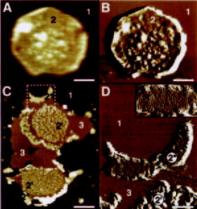
Height (A) and deflection (B) images of an intact disc (double-layered membrane). Two different surface types are discerned: mica (1) and the cytoplasmic surface of the disc (2). C, height image of open, flattened single-layered discs. Three additional surface types are visible: small raftlike structures (2′), larger paracrystalline Rho domains (2*), and lipid (3). The area marked by the broken white box is displayed in D as a deflection image at higher magnification; lattice lines from the Rho paracrystal (2*) are visible. Inset in D, magnification of the paracrystalline area (2*). Discs and disc membranes were adsorbed on mica and imaged in buffer solution at room temperature. Scale bars, 200 nm (A and B), 500 nm (C), and 150 nm (D). Frame size of the inset in D is 267 × 143 nm. Vertical brightness ranges are 30 nm (A), 0.6 nm (B), 18 nm (C), and 0.4 nm (D).