
Fig. 5. Chromatographic separation of nonpolar retinoids in the blood and liver from Lrat−/− mouse.
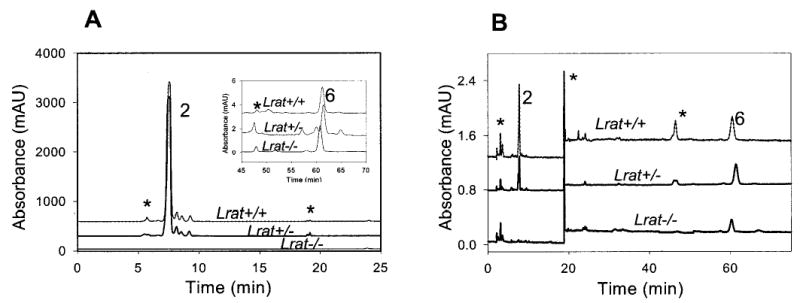
A, liver; B, blood. Retinoids were extracted from the tissues and separated on normal phase HPLC. The peaks correspond to the following retinoids: 2, all-trans-retinyl esters; 6, all-trans-retinol. * indicates artifacts related to a change in the solvent composition or compounds unrelated to retinoids.