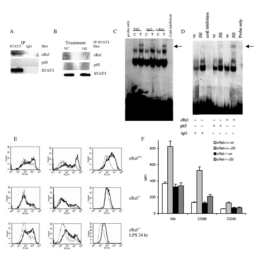
Figure 6.
Mechanism of JSI-124 induced activation of NF-κB in DCs. (A) CD11c± DCs were generated as described above and cells were stimulated with 20 ng/ml TNFα for 40 min. Immunoprecipitation was performed with anti-STAT3 antibody or control rabbit IgG as described in Material and Methods. Membranes were then probed with antibodies against cRel and STAT3. (B) CD11c± DCs treated with JSI-124 or DMSO (VC) for 36 hrs were collected, cell lysates were prepared. STAT3 was precipitated using anti-STAT3 antibody. Membranes were then probed with antibodies against cRel, p65, and STAT3. (C) CD11c±DCs were generated as described in Fig. 4 and treated for 20 min with 20 ng/ml TNFα (T). In control (C) cells were cultured in medium alone. Nuclear extracts were prepared and EMSA was performed with NF-κB specific probe in the presence of 5 μg of rabbit IgG or rabbit polyclonal antibodies against p65 or cRel (Santa Cruz). Arrow points on the place of supershift. Cold inhibition − EMSA performed in the presence of x100 excess of unlabeled probe. Probe only − EMSA performed without nucleoproteins. (D). CD11c±DCs were treated with either JSI-124 or DMSO (VC) for 48 hr. Nuclear extracts were prepared and EMSA was performed as described above. Arrow points on the place of supershift. Cold inhibition − EMSA performed in the presence of x100 excess of unlabeled probe. Probe only − EMSA performed without nucleoproteins. (E, F) DCs were generated from BM HPCs obtained from cRel-/- or control cRel+/+ mice. CD11c+ cells were isolated using magnetic beads separation technique and treated with 0.5 μM JSI-124 (solid line) or VC (dotted line) for 5 days. Cells then were collected and expression of MHC class II and co-stimulatory molecules was analyzed by flow cytometry. Typical example (E) and cumulative results (F) of three performed experiments are shown. In the bottom row labeled “cRel-/- LPS 24 hr”, DCs generated from cRel-/- HPCs were isolated using CD11c marker and were immediately activated for 24 hr with 5 μg/ml LPS. In that case solid line represent cells treated with LPS.

