
Fig. 2.
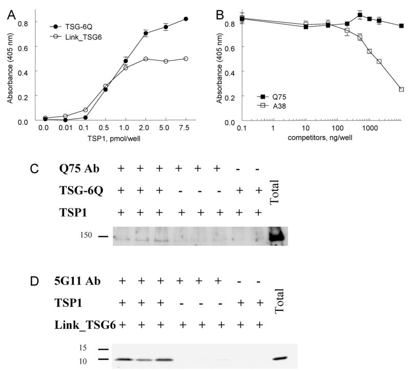
Binding of TSG-6 to immobilized TSP1. Panel A: Binding of 4 pmol solution phase TSG-6Q (filled circles) or 4 pmol Link_TSG6 (open circles) to the indicated amounts of surface bound TSP1 was determined by an ELISA using a rabbit polyclonal anti-TSG-6 antibody. Panel B: Interaction of solution phase TSG-6Q (4 pmol) with surface bound TSP1 (2 pmol) was assessed in the presence of the indicated concentrations of murine monoclonal TSG-6 antibodies Q75 (filled squares) or A38 (open squares). Absorbance at 405 nm is plotted as mean ± SEM, N = 8. Panel C: Binding of TSP1 to TSG-6Q in solution was detected by pull down using immobilized TSG-6 antibody Q75, which did not inhibit binding in B. Bound TSP1 was detected after solubilization in SDS, separation by SDS gel electrophoresis and blotting with biotinylated-TSP1 antibody A6.1, streptavidin-peroxidase, and ECL. Panel D: TSP1-biotinylated-Link_TSG6 complexes were pulled down using immobilized TSP1 antibody 5G11, which recognizes the third properdin repeat of TSP1 (99). Bound Link_TSG6 was detected by electrophoresis on 12% acrylamide Tris/HCl SDS gels and blotting using streptavidin-peroxidase and ECL.