
Fig. 5.
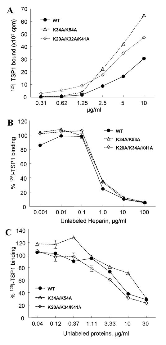
Inhibition of TSP1 binding to Link_TSG6 by heparin. A: Comparison of the TSP1-binding activities of Link module mutants with wild-type Link_TSG6. The binding of 125I-TSP1 to different amounts of wild-type (WT) Link_TSG6 (solid line) or selected mutants (dotted lines) was tested by solid phase assay. Background values are subtracted from each data point (256 ±14). B: The binding of 125I-TSP1 to different amounts of wild-type (WT) Link_TSG6 (solid line) or selected mutants (dotted lines) in the presence of the indicated concentrations of heparin. C: The binding of 125I-TSP1 to wells coated with heparin-BSA was determined in the presence of competing concentrations of unlabeled WT Link_TSG6 (solid line) or mutants (dotted lines). The assays were performed at 37°C, pH 7.3 in the presence of Ca2+, Mg2+. Results (mean cpm ± S.E.M.) shown are representative of two independent experiments performed in triplicate.