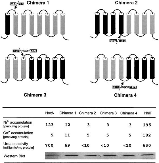
FIG. 2.
Structure and activity of chimeric nickel permeases. TMDs are shown as black (NhlF moiety) and grey (HoxN moiety) cylinders. The sequences at the fusion sites (arrows) in periplasmic (chimeras 1 and 3) and cytoplasmic (chimeras 2 and 4) loops are indicated in single-letter code. Construction of chimeras 3 and 4 resulted in four additional amino acid residues (PGDP) at the fusion sites. FLAG epitope-tagged chimeras 1 to 4 and parental transporters were individually produced in E. coli CC118. The metal accumulation of growing cells was assayed in Luria-Bertani medium supplemented with 500 nM concentrations of radiolabeled metal chlorides. The urease-enhancing activity of the permeases was analyzed in cells coexpressing a bacterial urease operon during growth in Luria-Bertani medium supplemented with 500 nM NiCl2. The relative quantities of the transporters were estimated by Western immunoblotting after separation of the solubilized membrane proteins by SDS-PAGE.

