
Figure 1.
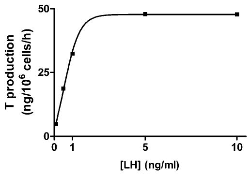
A representative concentration-response curve for luteinizing hormone (LH) in incubated rat Leydig cells. Purified rat Leydig cells were prepared as described in “Materials and Methods” and incubated in the presence of increasing concentrations of LH. Results are presented as nanograms of testosterone formed per 106 cells during a 1-hour incubation. This experiment was repeated 3 times with similar results.