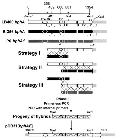
FIG. 1.
Strategies to evolve LB400 BphA. The constructions used for shuffling are drawn in the top part of the figure. These are LB400 and B-356 bphA cloned in pDB31 and P6 bphA1 cloned in pQE31. Large arrows represent genes; the gray box on the right side of the genes represents the vectors. Portions of LB400, B-356 bphA, or P6 bphA1 were PCR amplified with appropriate primers (indicated as small horizontal arrows) as described in the text. The appropriate DNA fragments for each of the three shuffling strategies were digested with DNase I and reassembled by primerless PCR. The primerless PCR products were amplified with internal primers (primers 6 or 7 and primer 8) to generate the libraries of MluI/AvrII hybrid fragments that were used to replace the corresponding fragment of LB400 bphA in pDB31[LB400-bphAE]. The numbers on top of the figure indicate the base pair positions in LB400 bphA. The indicated restriction sites are unique and common to both LB400 and B-356 bphA. Circles indicate the position of the 2Fe-2S Rieske center and the mononuclear Fe+ active center. I, II, III, and IV refer to the localization of the designated bphA regions I, II, III, and IV, respectively, of Mondello et al. (27).

